
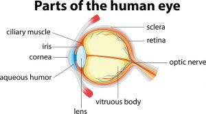

In healthy individuals, light travels through the cornea past the lens to the retina, allowing the brain to create an image. The corneal surface is normally smooth, so light can pass through with ease. In case of keratoconus, the cornea becomes cone-shaped and its surface irregular, so the passing light is projected as a distorted image.
Here we will further explain the causes, symptoms, and treatment of keratoconus.
Keratoconus causes
We know that in keratoconus the cornea becomes cone-shaped, but how exactly does this occur? The cornea is held in place by tiny fibers of the protein collagen. When these fibers weaken, the shape of the cornea can no longer be sustained and changes over time.
Keratoconus is caused by the decrease in protective antioxidants in the cornea. Antioxidants protect the fibers that hold the cornea in place, but if antioxidant levels are low, there is not enough protection available, hence the gradual weakening of collagen fibers.
It is quite common to see keratoconus run in families, so if parents or grandparents have had the condition, it’s important for children to monitor their eye health regularly. Keratoconus can take place as early as teenage years, and the shape of the cornea can change either rapidly or slowly.
Keratoconus: Signs and symptoms


In its early stage, keratoconus can be difficult to diagnose, as it can resemble other common eye problems. Proper diagnosis of keratoconus involves a trained eye professional.
Keratoconus and vision loss
The changes in the cornea associated with keratoconus severely impair the vision. In worst cases, a corneal transplant is required to restore vision. Laser eye surgery is not recommended for those living with keratoconus, because it can further weaken the cornea and lead to vision loss.
Keratoconus diagnosis

Your doctor will also need to measure the shape of the cornea to confirm that the physical change has taken place.
Keratoconus treatment
Early treatment requires the use of prescription glasses or rigid contact lenses. There is a specific procedure known as PTK that helps smooth out the scar and improve contact lens wear.
Another treatment is known as collagen crosslinking, which is effective for preventing keratoconus progression. Implants called intacs are placed beneath the surface of the cornea to help reduce the cone shape and improve vision.
The last resort is cornea transplant, which replaces the patient’s cornea with a donor cornea that is stitched into place.
The type of treatment depends on the severity of keratoconus.