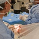
 Detached retina – retinal detachment – occurs when the retina moves away from its normal position. This can be a result of a tear, break, or hole. When the vitreous gel pulls loose – this is a clear gel inside of the eye and in front of the retina – it can exert traction on the retina. If the retina is already weak, it will tear and ultimately detach.
Once the retina detaches, the liquid from the vitreous gel starts accumulating behind the retina, further causing the retina to separate, leading to complete retinal detachment.
Advertisement
It is quite rare that retinal detachment occurs in both eyes, as it commonly affects one eye at a time. If retina detachment does occur in one eye, it is imperative to constantly monitor the other eye for any signs of possible retinal detachment.
When the retina becomes detached, it can no longer receive blood and nutrients, so the longer it goes untreated the greater the risk for permanent vision loss is.
Detached retina causes and risk factors
A detached retina is not commonly caused by injury, but an injury could result in retinal detachment. Other possible causes for retinal detachment include a sagging vitreous and advanced diabetes.
Age can be a factor in retinal detachment, too. With aging, the thinning retina becomes more prone to tears, or the changes to the vitreous gel result in fluid accumulation under the retina, pulling it away from the underlying tissues and cutting the blood supply in these areas.
Other risk factors for retinal detachment include a previous retinal detachment, a family history of retinal detachment, extreme nearsightedness, a previous eye injury, and any other eye disease or inflammation.
Detached retina symptoms
 Although you won’t necessarily feel anything when your retina starts detaching, there are early warning signs to watch out for so you can receive treatment sooner rather than later when the risk of complications arise.
Signs and symptoms of retinal detachment include:
- Sudden appearance of floaters
- Flashes of light
- Blurred vision
- Gradually reduced peripheral vision
- Curtain-like shadow over the visual field
You should see a doctor for retinal detachment if you begin to experience any of the above symptoms. Furthermore, if you are over the age of 50, if you have had a family member with retinal detachment, and if you’re extremely nearsighted, you should see a doctor right away.
Retinal detachment diagnosis
Your doctor will perform a retinal examination and ultrasound imaging in order to properly diagnose retinal detachment. During a retinal examination, the doctor uses an instrument that produces a bright light and a special lens to examine the back of the eye. Ultrasound imaging is used to look past any bleeding that may have occurred preventing the doctor from seeing the retina.
Treatment for retinal detachment
 Surgery is the main treatment when dealing with any tears, holes, or retinal detachment. For retinal tears, laser surgery or freezing are used as a means to heal the tear or hole. These types of procedures are usually done on an outpatient basis, so you can go home soon after they are done. You may have to modify your activities while recovering as per doctor’s instructions.
To treat retinal detachment, the surgeon may inject air or gas into the eye, creating a bubble that pushes the hole against the retina and stops the flow of fluid into the open space.
Advertisement
Another treatment is indenting the surface of the eye, which basically involves sewing a silicone patch to the white of the eye, indenting the wall of the eye in order to relieve pressure.
Lastly, the doctor can drain the excess fluid from the eye and instead inject either gas, air, or silicone into the vitreous space to help flatten the retina.
Depending on your condition, your doctor will decide which mode of treatment is best.

